

![]() ✍️ B-scan produces two-dimensional images.
✍️ B-scan produces two-dimensional images.
![]() ✍️ shifting the probe and the patient’s gaze to bring lesions into the central, most sensitive part of the beam path to produce best echograms
✍️ shifting the probe and the patient’s gaze to bring lesions into the central, most sensitive part of the beam path to produce best echograms
![]() ✍️ Placing lesions in the center of the echogram by allowing the patient to direct their gaze to the desired position
✍️ Placing lesions in the center of the echogram by allowing the patient to direct their gaze to the desired position
![]() ✍️ applying the probe directly on the globe through open lids.
✍️ applying the probe directly on the globe through open lids.
![]() ✍️ scanning through open lids also improves the resolution and quality of images by minimizing sound attenuation from the lids.
✍️ scanning through open lids also improves the resolution and quality of images by minimizing sound attenuation from the lids.
 ✍️ The majority of patients do scanning directly on the anesthetized globe
![]() 👉 traumatized eyes
👉 traumatized eyes
![]() 👉 in children
👉 in children
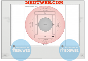
✅ B-scan orientation and sections
![]() ✍️ a marker (placed near the probe face )determines the axis of the beam on the fundus and indicates the upper end of the echogram on the display screen
✍️ a marker (placed near the probe face )determines the axis of the beam on the fundus and indicates the upper end of the echogram on the display screen
![]() ✍️ the optic nerve is used as an echographic center of the globe and as a reference
✍️ the optic nerve is used as an echographic center of the globe and as a reference
✍️ the globe is divided concentrically into three equal zones
![]() 👉 posterior (P)
👉 posterior (P)
![]() 👉 equator (E)
👉 equator (E)
![]() 👉 anterior (A)
👉 anterior (A)
![]() ✍️ the globe is further divided radially into clock hour meridians
✍️ the globe is further divided radially into clock hour meridians
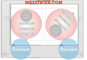
✅B-scan echographic sections
✍️ Axial section
![]() 👉the patient fixates at the primary gaze and the probe is placed on the cornea and directed axially.
👉the patient fixates at the primary gaze and the probe is placed on the cornea and directed axially.
![]() 👉 Sections of all the clock hours can be performed by rotating the probe on its axis
👉 Sections of all the clock hours can be performed by rotating the probe on its axis
![]() 👉 the echograms are labeled after the location and orientation of the beam on the fundus
👉 the echograms are labeled after the location and orientation of the beam on the fundus
![]() 💧 horizontal axial
💧 horizontal axial
![]() 💧 vertical axial
💧 vertical axial
![]() 💧 1.30 axial
💧 1.30 axial
![]() 💧 10.30 axial
💧 10.30 axial
![]() 👉 the posterior lens surface and optic nerve head are placed in the center of the echogram
👉 the posterior lens surface and optic nerve head are placed in the center of the echogram
✍️ Longitudinal section
![]() 👉 along one meridian only, from the optic nerve (lower portion of the echogram) to the ciliary body (upper portion of the echogram)
👉 along one meridian only, from the optic nerve (lower portion of the echogram) to the ciliary body (upper portion of the echogram)
![]() 👉 created by placing the probe on the sclera near the limbus with the marker radially placed at its corneal side(avoiding scanning through the lens).
👉 created by placing the probe on the sclera near the limbus with the marker radially placed at its corneal side(avoiding scanning through the lens).
![]() 👉 the periphery and ciliary body are brought into view by directing the patient’s gaze 180° away from the probe marker (toward the meridian to be scanned)
👉 the periphery and ciliary body are brought into view by directing the patient’s gaze 180° away from the probe marker (toward the meridian to be scanned)
![]() 👉 the echograms are labeled after the clock hour location of the beam (not the probe and marker).
👉 the echograms are labeled after the clock hour location of the beam (not the probe and marker).
![]() 💧L12 section is created by a probe and marker placed at 6 o′clock
💧L12 section is created by a probe and marker placed at 6 o′clock
![]() 💧 L1:30 by a probe and marker placed at 7:30
💧 L1:30 by a probe and marker placed at 7:30
✍️ Transverse section
![]() 👉 the probe is placed on the scleral side of the limbus and directed posteriorly.
👉 the probe is placed on the scleral side of the limbus and directed posteriorly.
![]() 👉 single smooth arc movement (following the curvature of the globe) it is shifted and rotated anterior–posteriorly, from the limbus to the fornix
👉 single smooth arc movement (following the curvature of the globe) it is shifted and rotated anterior–posteriorly, from the limbus to the fornix
![]() 👉 scanning the opposite globe wall posterior–anteriorly.
👉 scanning the opposite globe wall posterior–anteriorly.
![]() 👉 Perpendicularity is helped by directing the patient’s gaze 180° away from the probe.
👉 Perpendicularity is helped by directing the patient’s gaze 180° away from the probe.
![]() 👉 the echograms are labeled according to the clock hour at the center of the beam, and also to the beam’s anterior–posterior location.
👉 the echograms are labeled according to the clock hour at the center of the beam, and also to the beam’s anterior–posterior location.
![]() 💧 a section labeled transverse 12 posterior (12P) is produced by a probe located at 6 o′clock limbus
💧 a section labeled transverse 12 posterior (12P) is produced by a probe located at 6 o′clock limbus
![]() 💧 transverse 7:30 posterior (P), equator (E), anterior (A) sections are produced by placing the probe at 1:30 limbus, mid distance, and fornix respectively
💧 transverse 7:30 posterior (P), equator (E), anterior (A) sections are produced by placing the probe at 1:30 limbus, mid distance, and fornix respectively
![]() 👉 in transverse sections, it is conventional to place the probe marker nasally instead of temporally, and up instead of down.
👉 in transverse sections, it is conventional to place the probe marker nasally instead of temporally, and up instead of down.
✅B-scan Protocol for screening of the globe with B-scan
![]() ✍️ Topical anesthetic is instilled into the eye.
✍️ Topical anesthetic is instilled into the eye.
![]() ✍️ Examination is conducted through open lids, directly on the globe
✍️ Examination is conducted through open lids, directly on the globe
![]() ✍️ Coupling jelly(methylcellulose) is applied onto the tip of the probe
✍️ Coupling jelly(methylcellulose) is applied onto the tip of the probe
✍️ Four transverse sections are performed as follows
![]() 👉 Transverse 12
👉 Transverse 12
![]() 💧 the patient looks up at 12 o’clock
💧 the patient looks up at 12 o’clock
![]() 💧 the probe is placed at the 6 o′clock limbus with its marker nasally.
💧 the probe is placed at the 6 o′clock limbus with its marker nasally.
![]() 💧 Shifting and rotating the probe from limbus to fornix and scanning the superior retina posterior–anteriorly
💧 Shifting and rotating the probe from limbus to fornix and scanning the superior retina posterior–anteriorly
![]() 👉 Transverse 3
👉 Transverse 3
![]() 💧the patient looks toward 3 o′clock
💧the patient looks toward 3 o′clock
![]() 💧the probe is placed at the 9 o′clock limbus with its marker up.
💧the probe is placed at the 9 o′clock limbus with its marker up.
![]() 💧 the probe is shifted and rotated from limbus to fornix, scanning the nasal retina in the right eye and temporal retina in the left eye posterior–anteriorly
💧 the probe is shifted and rotated from limbus to fornix, scanning the nasal retina in the right eye and temporal retina in the left eye posterior–anteriorly
![]() 👉 Transverse 6
👉 Transverse 6
![]() 💧the patient looks down at 6
💧the patient looks down at 6
![]() 💧 the probe is placed at the 12 o′clock limbus with its marker nasally.
💧 the probe is placed at the 12 o′clock limbus with its marker nasally.
![]() 💧 Shifting and rotating the probe from limbus to fornix and scanning the inferior retina posterior–anteriorly
💧 Shifting and rotating the probe from limbus to fornix and scanning the inferior retina posterior–anteriorly
![]() 👉 Transverse 9
👉 Transverse 9
![]() 💧 The patient looks toward 9 o′clock
💧 The patient looks toward 9 o′clock
![]() 💧 the probe is placed at the 3 o′clock limbus, with its marker up.
💧 the probe is placed at the 3 o′clock limbus, with its marker up.
![]() 💧 the probe is shifted and rotated from limbus to fornix, scanning the temporal retina in the right eye and nasal retina in the left eye posterior–anteriorly
💧 the probe is shifted and rotated from limbus to fornix, scanning the temporal retina in the right eye and nasal retina in the left eye posterior–anteriorly
![]() ✍️ Additional transverse scans of any of the other clock hour meridians (1:30, 4:30, 7:30, and 10:30)may be performed in a similar fashion, if an abnormality is suspected at the given meridian.
✍️ Additional transverse scans of any of the other clock hour meridians (1:30, 4:30, 7:30, and 10:30)may be performed in a similar fashion, if an abnormality is suspected at the given meridian.
![]() ✍️ the aim is always to place lesions in the center of the echogram
✍️ the aim is always to place lesions in the center of the echogram
![]() ✍️ Scans are performed first with a high gain to detect low reflective echoes (vitreous opacities)
✍️ Scans are performed first with a high gain to detect low reflective echoes (vitreous opacities)
![]() ✍️ repeat scans with a lower gain to achieve higher resolution (for better resolution of layers of the globe wall and accurate measurement of mass dimensions).
✍️ repeat scans with a lower gain to achieve higher resolution (for better resolution of layers of the globe wall and accurate measurement of mass dimensions).
![]() ✍️ If no abnormalities are found after employing the above protocol, no further examination is usually required.
✍️ If no abnormalities are found after employing the above protocol, no further examination is usually required.
![]() ✍️ If a lesion is detected, longitudinal and axial sections are additionally performed.
✍️ If a lesion is detected, longitudinal and axial sections are additionally performed.
![]() ✍️ Multiple sections are helpful for viewing lesions at different angles locating their clock hour meridian and anterior–posterior position and creating the three-dimensional impression.
✍️ Multiple sections are helpful for viewing lesions at different angles locating their clock hour meridian and anterior–posterior position and creating the three-dimensional impression.